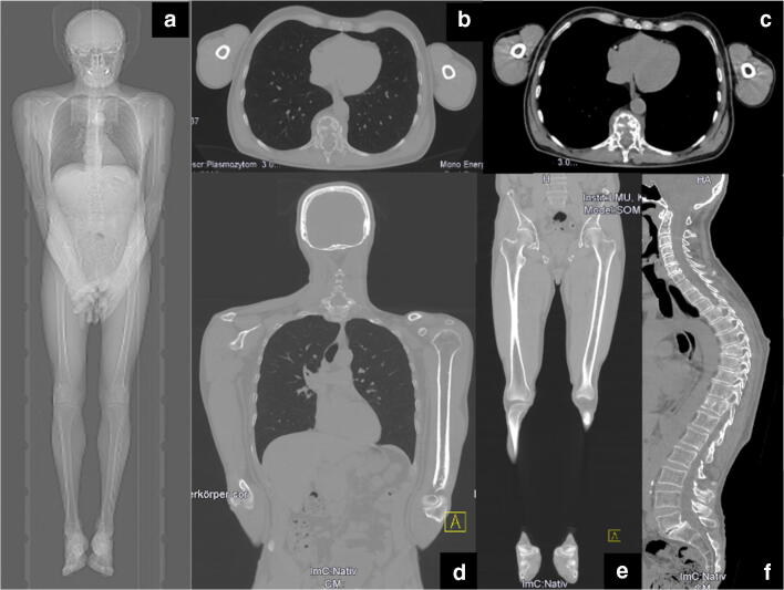
Fig. 2.
Image examples of a whole-body low-dose CT scan (a: scout). Images were reconstructed in axial 3 mm slices using the intermediate (b) and soft-tissue kernel (c) in order to evaluate soft-tissue masses and the intrathoracic and intra-abdominal organs. The skeleton is further reconstructed in coronal 3 mm slices using the bone kernel for the reading of the upper (skull, thorax, arms; b, d) and the lower parts (pelvis, legs; e) of the body. The spine is additionally reconstructed in sagittal orientation (f).

