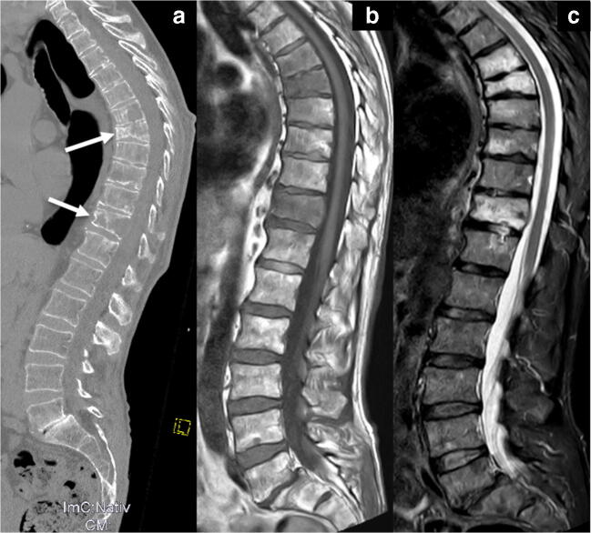
Fig. 3.
(a) Marked osteolytic lesions are detected in the 6th, 7th and 11th thoracic vertebral body and pathological fractures in the 7th and 11th vertebrae in the sagittal CT reconstruction of the spine in the bone window. Additional lesions are suspected in the 8th–10th vertebral body. MRI of the patient using a T1-weighted (b) and a STIR (c) sequence in sagittal orientation confirms the involvement of myeloma in the 6th, 7th and 11th vertebrae, while end plate-associated edema due to degenerative disease is seen in the 8th and 9th thoracic vertebrae. Thus, CT can sometimes over-diagnose myeloma involvement in cases of osteoporosis and inhomogeneous bone structure

