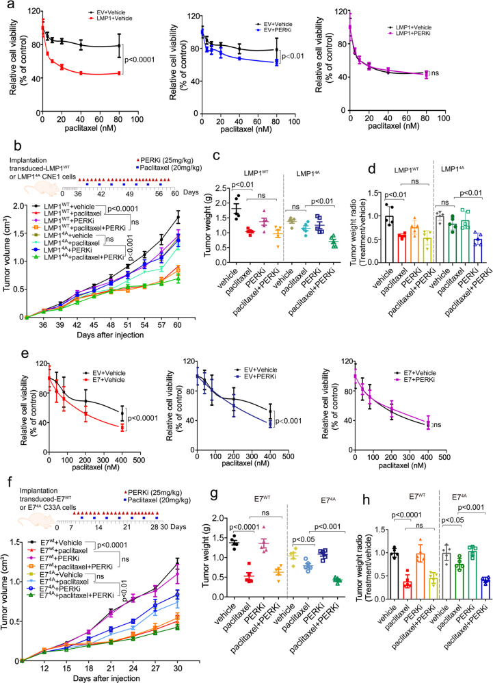
Fig. 8.
PERK inhibition by DNA tumor virus oncogenes increases cancer cell chemosensitivity. a, e CNE1-EV or CNE1-LMP1 (a) and C33A-EV or C33A-E7 cells (e) were pretreated with a vehicle or PERK inhibitor (PERKi) for 24 h, followed by treatment with paclitaxel at the indicated doses for 48 h. Cell viability was measured using a CCK8 assay. Data are presented as the mean ± SEM. b–d Tumor growth by 3 × 10⁶ subcutaneously injected CNE1 cells stably expressing LMP1WT or LMP14A. b CNE1 cells stably expressing LMP1WT or LMP14A (3 × 10⁶) were injected into nude mice. Once the tumor volume reached 100–120 mm3, the mice were treated once daily with a vehicle or PERKi (25 mg/kg) by oral administration and twice weekly with paclitaxel (20 mg/kg) or PBS by intraperitoneal injection according to the schedule described in the “Materials and methods”. The tumors were collected on day 60 and tumor weight (c) was quantified. The raw data are shown in (c), and normalized data are shown in (d). f–h A total of 3 × 10⁶ C33A cells stably expressing E7WT or 6 × 10⁶ C33A cells stably transfected with E74A were transplanted into nude mice (f). Once the tumor volume reached 60–80 mm3. The mice were treated as described above, except the tumors were collected on day 30. Tumor weight was quantified and is shown in (g); normalized data are shown in (h). n = 5 mice per group, the data are presented as the mean ± SEM. The values of p < 0.05 were considered statistically significant. ns means no significant

