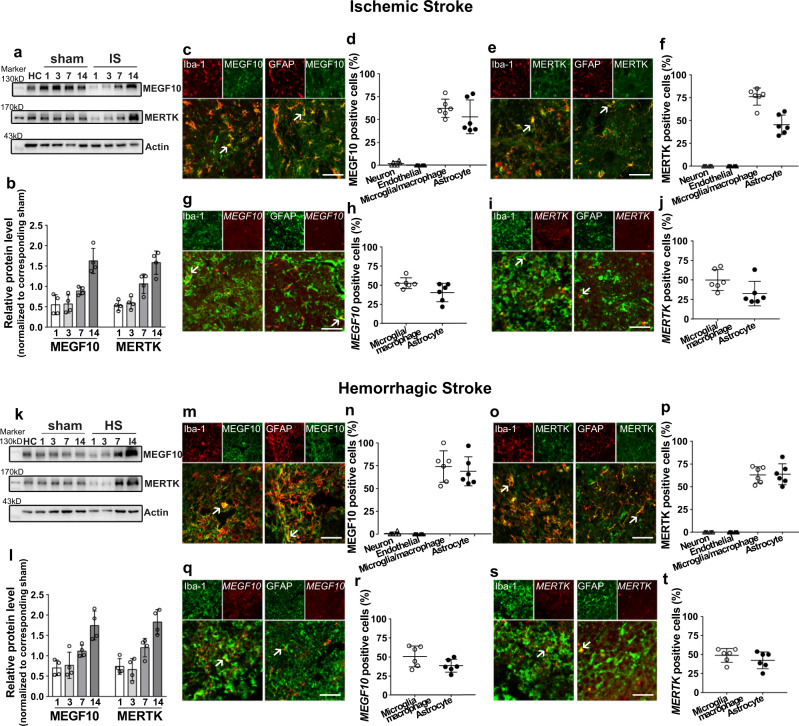
Fig. 4. Upregulation of MEGF10 and MERTK levels in mouse brain following ischemic and hemorrhagic stroke.
Western blotting and quantification of MEGF10 and MERTK protein levels (normalized to corresponding sham) in ischemic (a) and hemorrhagic mouse brain (k). Immunostaining showed MEGF10 (green) and MERTK (green) were expressed in Iba-1+ microglia/macrophages (red) and GFAP+ astrocytes (red) in ischemic (c–f) and hemorrhagic stroke (m–p). Arrows indicated colocalization of MEGF10 and MERTK with microglia/macrophages and astrocytes. Bar = 50 µm. Fluorescence in situ hybridization study showed MEGF10 (red) and MERTK (red) signals colocalized in Iba-1+ microglia/macrophages (green) and GFAP+ astrocytes (green) in ischemic (g–j) and hemorrhagic stroke (q–t), indicated by arrows. Bar = 50 µm. (b, l) N = 4 mice per group. (d, f, h, j, n, p, r, t) Statistics are derived from six slices, n = 3 mice per group. Data are mean ± SD.

