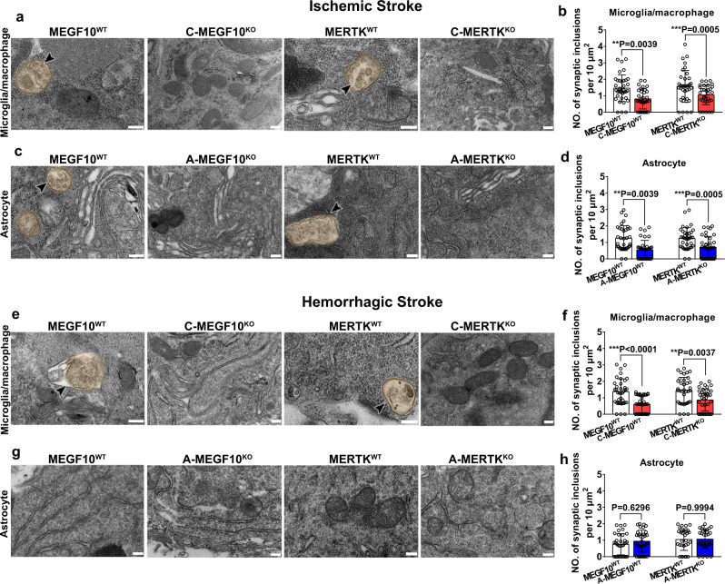
Fig. 6. Conditional MEGF10 or MERTK knockout reduced glial cell-mediated synapse engulfment after stroke.
a–d TEM images showed Iba-1+ microglia/macrophages and GFAP+ astrocytes in the gliosis region enwrapped engulfed synaptic elements in MEGF10WT and MERTKWT mice, but the number of engulfed synaptic elements were reduced in C-MEGF10KO, C-MERTKKO, A-MEGF10KO, and A-MERTKKO mice after ischemic stroke. Arrowheads indicate synaptic elements that were engulfed by microglia/macrophages (a, b) or astrocytes (c, d). Statistics are derived from 36, 32, 36, 31 cells (b) and 36, 36, 36, 36 cells (d) (from left to right), n = 3 mice per group. e–h TEM images showed Iba-1+ microglia/macrophages in the gliosis region contained synaptic elements in MEGF10WT and MERTKWT mice (e, f); while synaptic elements were rarely detected in GFAP+ astrocytes (g, h). Statistics are derived from 36, 36, 36, 36 cells (f) and 34, 34, 30, 34 cells (h) (from left to right), n = 3 mice per group. Bar = 200 nm. One-way ANOVA followed by Tukey’s test. Data are mean ± SD.

