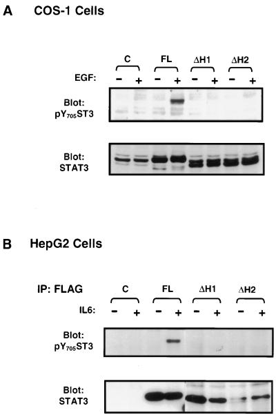
FIG. 3.
Deletion of helix α1 or α2 abolishes tyrosine phosphorylation of Stat3 in response to EGF or IL-6. COS-1 (A) or HepG2 (B) cells were transfected with the control plasmid (C), full-length Stat3 (FL), or the mutants in which either helix α1 (ΔH1) or α2 (ΔH2) was deleted and either left untreated (−) or treated with EGF or IL-6 (+). The tyrosine phosphorylation and expression of Stat3 were monitored by Western blot analysis (A) and immunoprecipitation/blotting analysis (B,) as described in the legend to Fig. 1.

