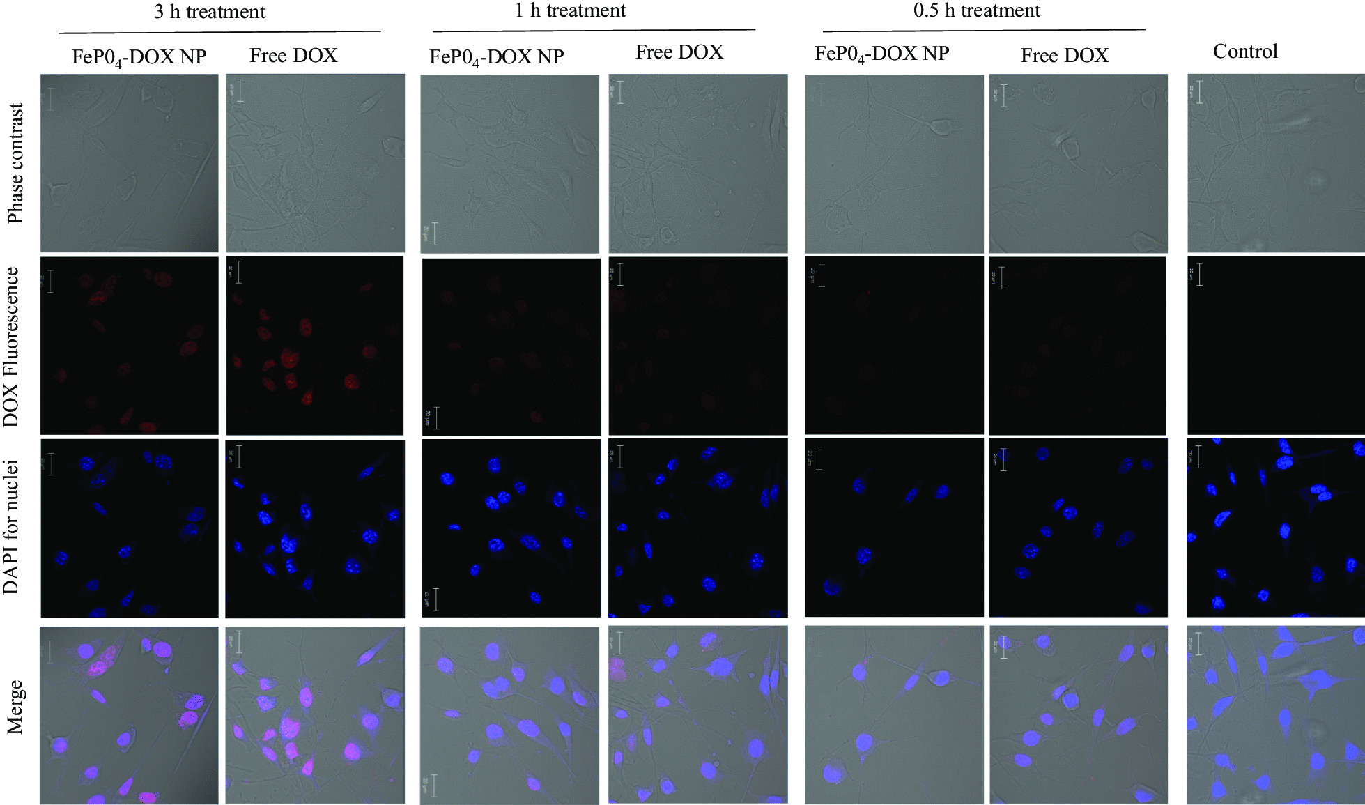
Fig. 4.

Cellular internalization study. Cellular internalization of FePO4-DOX NPs and Free DOX on K7M2 cells after 3 h, 1 h, and 0.5 h treatment. Cells were treated with 200 µL of 5 µg/mL DOX concentration. The red color observed in nanoparticle treated cell line signify successful internalization of the nanoparticles. The red color is due to the fluorescence characteristic of DOX. No red signal is observed in the untreated control cell
