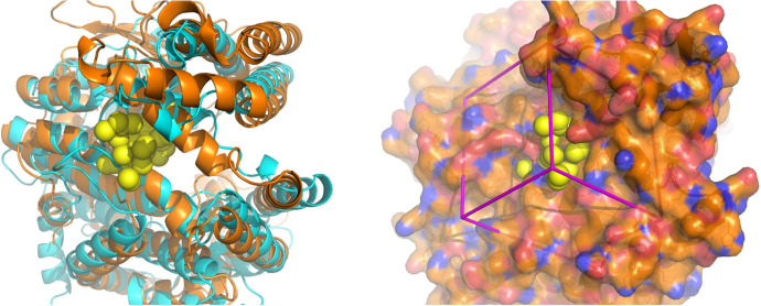
Fig. 1.
Surface pocket in the hinge region selected for molecular docking. Left panel: superposition of ribbon diagrams of ACE2 (cyan) and Nln (orange) and their hinge regions. Crystal structures of the open conformations of both peptidases are shown (Brown et al., 2001; Towler et al., 2004). The yellow spheres represent the site in the hinge region of Nln selected for molecular docking. Right panel: the crystal structure of Nln shown in the same orientation as in the left panel. The molecular surface is colored gold for carbon, red for oxygen, and blue for nitrogen. Yellow spheres depict sites for potential ligand atoms used in molecular docking. The box in magenta represents the boundaries of the scoring grid used to generate scores that consider electrostatic (polar) and van der Waals (nonpolar) interactions.

