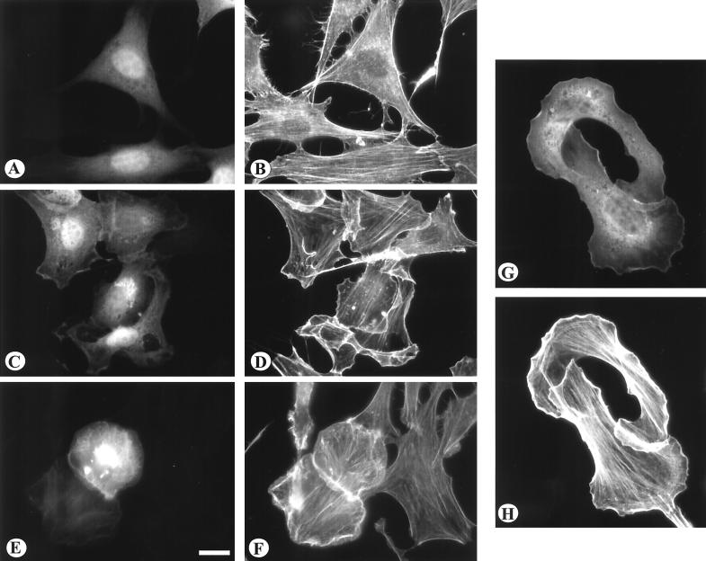
FIG. 3.
The effects of Vav2 transient expression on the actin cytoskeleton. NIH 3T3 cells (A to F) were transiently transfected with GFP alone (A and B), with wild-type Vav2 as a GFP construct (C and D), or with the Δ184N Vav2-GFP (E and F). BALB/c3T3 cells (G and H) were transfected with Δ184N Vav2 as a GFP fusion protein. Transfected cells were visualized for GFP (A, C, E, and G). The distribution of actin was visualized by staining with phalloidin conjugated with Texas red (B, D, F, and H). Note that expression of both wild-type and Δ184N Vav2 induced prominent lamellipodia and membrane ruffles. Bar = 20μm.

