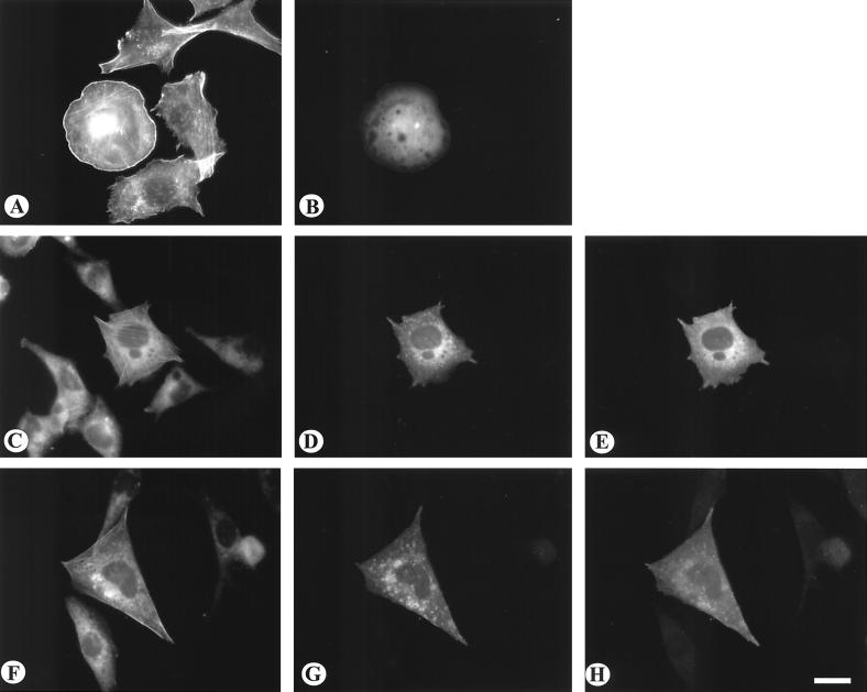
FIG. 6.
Effect of coexpressing dominant negative Cdc42 and Rac1 on the morphology of cells expressing constitutively active Vav2. CHO cells were transfected with Δ184N Vav2-GFP either alone (A and B) or cotransfected with myc-tagged N17Cdc42 (C to E) or myc-tagged N17Rac1 (F to H). Actin was visualized by staining with rhodamine-phalloidin (A, C, and F). Cells expressing Δ184N Vav2-GFP were visualized by GFP fluorescence (B, D, and G). Cells expressing the myc-tagged constructs were visualized by immunofluorescence (E and H). Bar = 20 μm.

