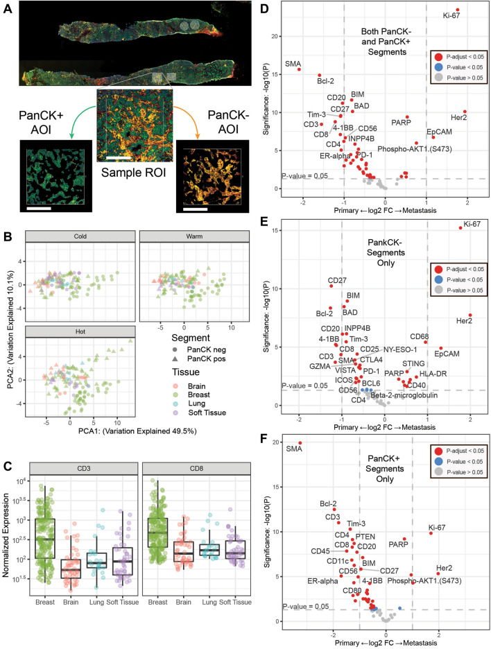
Fig. 2.
Digital spatial profiling of primary and metastatic tumors. A Visualization markers for Pan-Cytokeratin (PanCK, tumor—green), CD45 (total immune—red) and CD3 (T cells—yellow). Each ROI segmented tumor (PanCK +) or stroma (PanCK-) areas of illumination (AOI). Scale bar (white) is 250 microns. B Normalized Protein expression of CD3 and CD8 in primary and each metastatic site (lung, brain and soft tissue). C Principle components analysis of the 30 proteins with the highest coefficient of variation faceted by primary vs metastatic site and immune status (cold, warm and hot). D Differential expression of proteins from all ROI types and segments comparing primary breast and all metastatic. E Differential expression of proteins from all ROI types from the PanCK negative stromal segments comparing primary breast and all metastatic. F Differential expression of proteins from all ROI types from the PanCK positive tumor segments comparing primary breast and all metastatic. For D-F vertical dotted lines represent a onefold log2 change and the horizontal line marks an unadjusted p-value of p < 0.05. Dots in grey are not significant, dots in blue have an unadjusted p-value of < 0.05 and dots in red have an adjusted p-value of < 0.05

