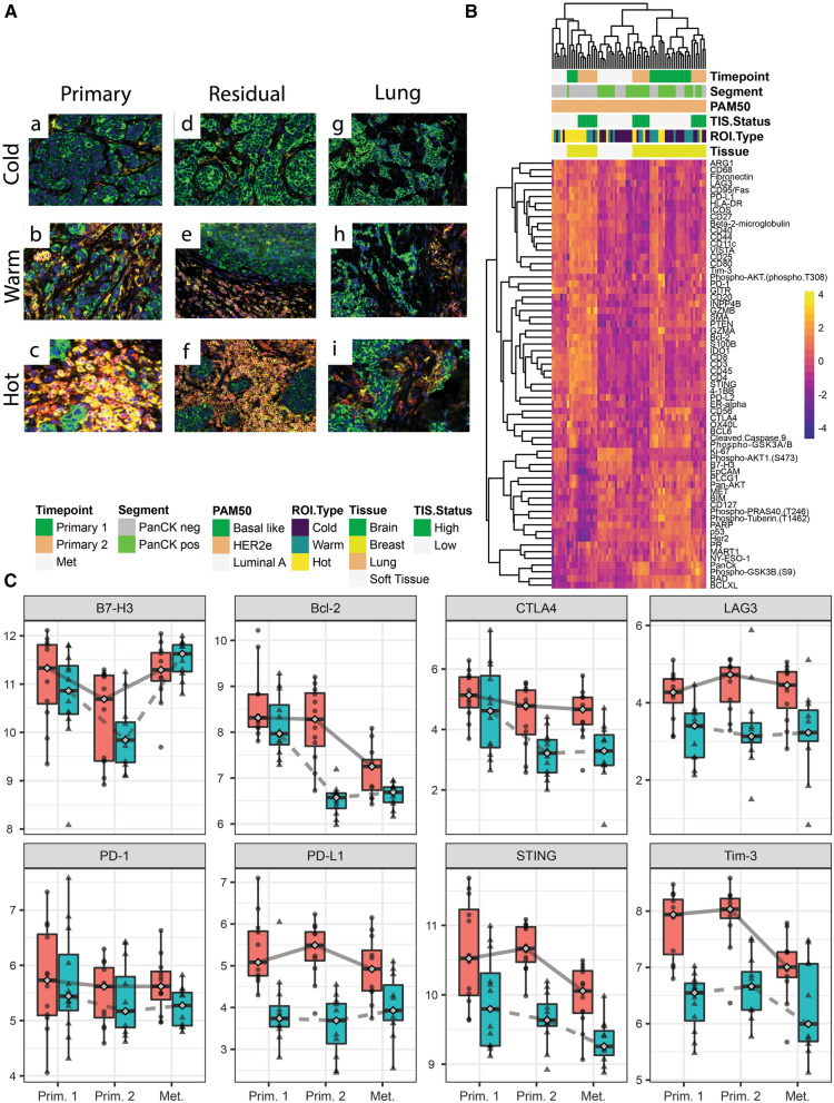
Fig. 4.
Analysis of primary, residual breast tumor and metastatic samples of patient 2. A Shows immune cold, warm and hot regions of interest for the three timepoints for patient 2 at primary, residual breast tumor and metastatic stages. Pan-cytokeratin is stained in green, CD45 in red and CD3 in yellow. B Unsupervised hierarchical clustering showing differences in protein expression profiling for each timepoint for patient 2. C Longitudinal plots showing log2 normalized protein expression for B7-H3, Bcl-2, CTLA4, LAG2, Tim3, PD1, PD-L1 and STING. The red boxplots with circles are PanCK− and blue boxplots with triangles are PanCK+ segment

