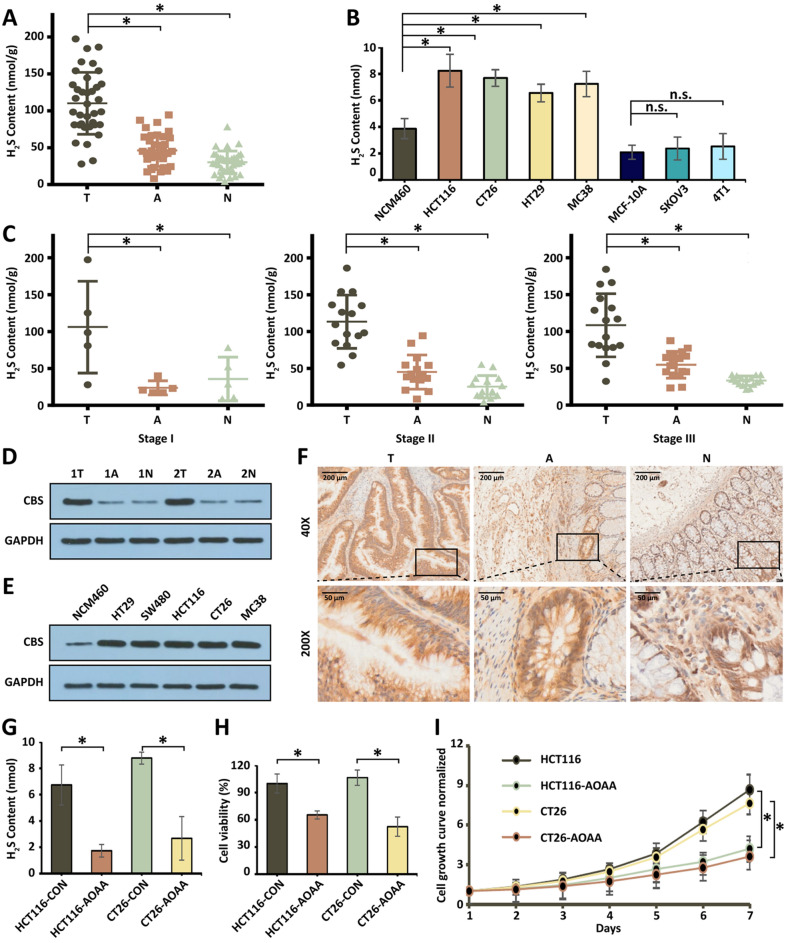
Fig. 1.
The overproduction of H2S in human CRC. A, B H2S production measured in human colorectal cancer specimens (*P < 0.05 vs. normal mucosa) and in colorectal cancer cell lines (*P < 0.05 vs. NCM460 cells; n.s.: no significant difference v.s. MCF-10A) by the H2S microsensor. (T: tumor tissue; A: aside colorectal tumor tissue; N: normal colorectal tissue). C H2S production in different stage of human colorectal cancer specimens measured by the H2S microsensor. D Western Blot of CBS protein expression in human colorectal cancer specimens, paired with the adjacent tissue and corresponding normal mucosa tissues. Membranes are re-probed for GAPDH expression to show that similar amounts of protein were loaded in each lane. E Immunohistochemistry of CBS protein expression in human colorectal cancer specimens, paired with the adjacent tissue and corresponding normal mucosa tissues. F Western Blot of CBS protein expression in colon cancer cell lines (vs. NCM460). G H2S production is measured in colon cancer cell lines treated by AOAA. H Cell viability of the HCT116 and CT26 treated with AOAA for 24 h. I Cell growth of the HCT116 and CT26 treated with AOAA within 6 days. (*P < 0.05), (n.s.: No significant difference, P value > 0.05)

