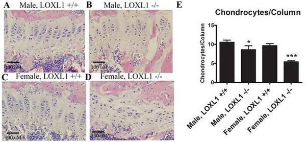
Figure 8. Hematoxylin and eosin staining of representative sagittal sections of femur distal growth plates.
(A) Images are from wild type and LOXL1−/− male and female mice; scale bar, 100 μM. (B) Histomorphometric analyses of histological sections of femur sections for chondrocytes per column of wild type and mutant male and female mice. Asterisk notation is the same as in Figure 6, n=8.

