
Fig. 2 |. Detection of mutations in liquid biopsy samples.
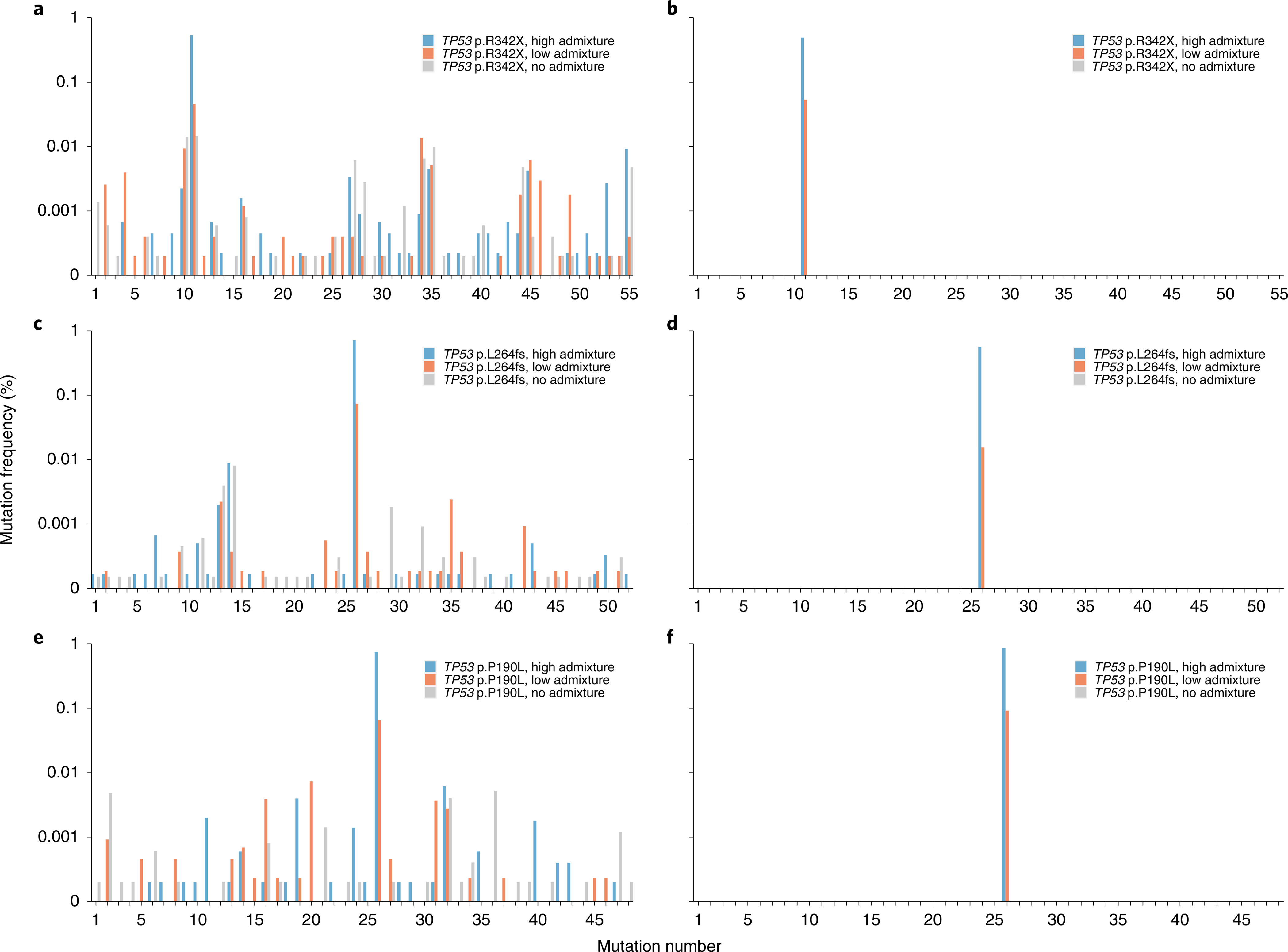
Analysis of 33 ng of plasma cell-free DNA from healthy individuals admixed with cell-free plasma DNA from an individual with cancer. Mixtures were created to generate a high frequency (~0.5–1%) of mutation (blue bars), low frequency (~0.01–0.1%) of mutation (orange bars) or no mutation (gray bars). The admixed TP53 p.R342X sample was assayed with SafeSeqs (a) and SaferSeqS (b). Similarly, the admixed TP53 p.L264fs sample was assayed with SafeSeqs (c) and SaferSeqS (d), and the admixed TP53 p.P190L sample was assayed with SafeSeqs (e) and SaferSeqS (f). Mutation numbers represent each of the 153 distinct mutations observed with SafeSeqS defined in Supplementary Table 2.