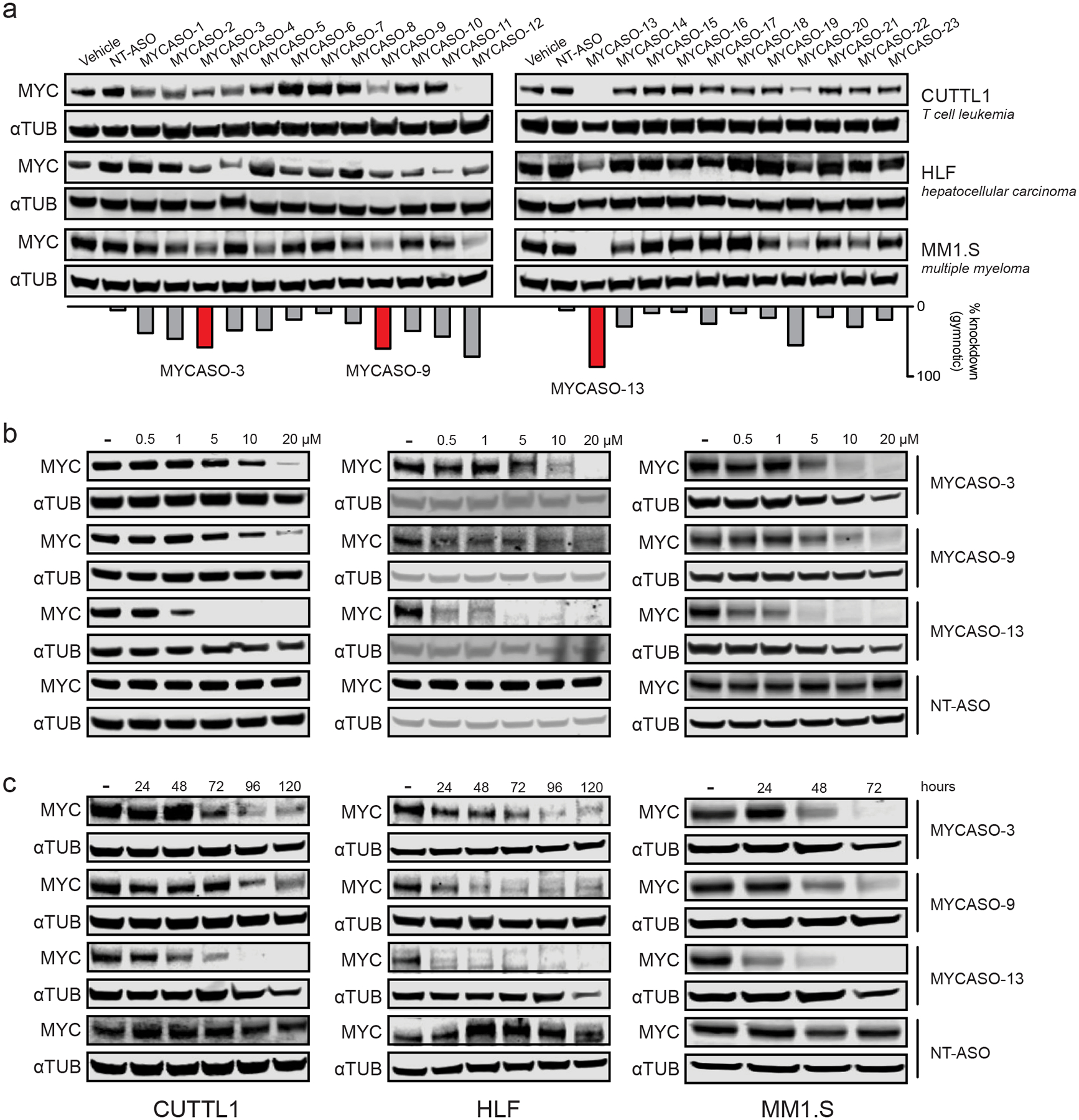
Figure 2: MYCASOs decrease MYC protein expression in cancer cell lines.

(a) Immunoblotting for MYC and alpha-tubulin in three MYC-expressing cancer cell lines after treatment with the full MYCASO library. Panels show CUTLL1 and MM1.S cells with 10 μM gymnotic-treated MYCASO for 72 hours. HLF cells were treated gymnotically at 10 μM for 120 hours. The bar graph shows average percent knockdown of MYC of gymnotic-treated cells across all four cell lines including HeLa. Highlighted MYCASOs (3, 9, and 13) were chosen for further study due to their superior knockdown activity and non-overlapping seed sites along the MYC mRNA. (b) Dose-proportional knockdown of MYC protein expression with MYCASO treatment. CUTLL1, MM1.S, and HLF cells were treated with 0, 0.5, 1, 5, 10, and 20 μM MYCASO (gymnotic) for 72 hours (CUTLL1 and MM1.S) or 120 hours (HLF). (c) Time-proportional knockdown of MYC protein expression with MYCASO treatment. CUTLL1 and HLF cells were treated with 10 μM MYCASO (gymnotic) for 24, 48, 72, 96, and 120 hours. MM1.S cells were treated with 10 μM MYCASO (gymnotic) for 24, 48, and 72 hours.
