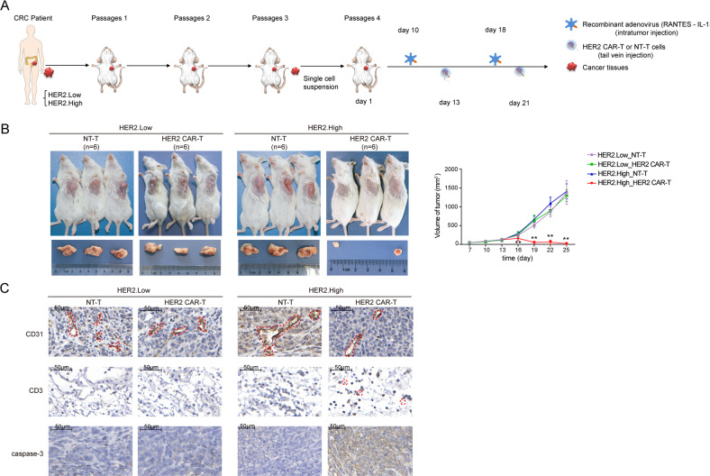
Fig. 5. HER2 CAR-T cells display greater aggressiveness in HER2+ CRC in PDX models.
A Left panel: Fresh human CRC tissues were transplanted into NCG mice. After three passages, a singlecell suspension of the PDX tumor cells (HER2.Low: low expression of HER2, HER2.High: high expression of HER2) was obtained. Right panel: Schematic of the PDX models (n = 24). NCG mice were injected subcutaneously with single-cell suspension on day 1, and recombinant adenovirus was injected on days 10 and 18. HER2 CAR-T or NT-T cells (1 × 10⁷) were injected via a tail vein on days 13 and 21, respectively. B The volume of the tumor was measured twice a week (**P < 0.01, HER2 CAR-T compared with NT-T in HER2.High group, two-tailed t-test). C The protein levels of CD31, CD3, and caspase-3 in mice were evaluated by IHC, and the representative images are shown. The scale bar is marked in each image.

