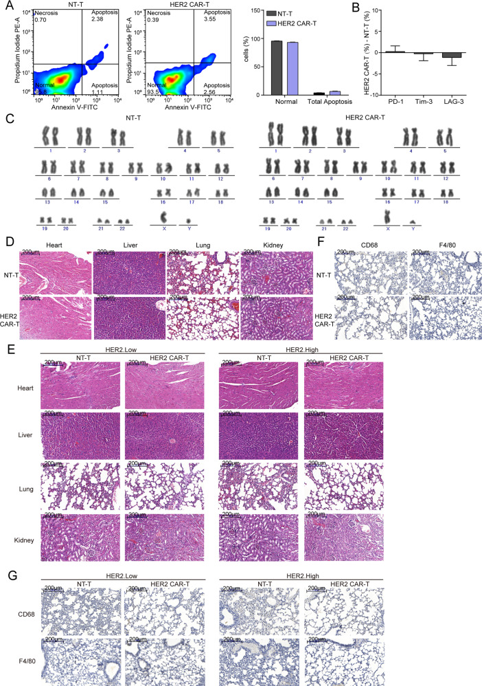
Fig. 7. Safety of HER2 CAR-T cells.
A 7 days after lentivirus infection, an apoptosis detection kit was used to detect the apoptosis of T cells, and the representative images are shown. B The difference in the expression levels of three exhaustion markers between HER2 CAR-T and NT-T group (HER2 CAR-T (%) - NT-T (%)) as detected by flow cytometry on 7 days after lentivirus infection. C 14 days after lentivirus infection, karyotype analysis was performed to detect chromosomal alterations in CAR-T cells. D, E H&E was performed to evaluate the pathological changes in Heart, Liver, Lung, and Kidney, and the representative images are shown. The scale bar is marked in each image. F, G IHC assay was performed to observe F4/80 or CD 68 positive macrophages in murine lung tissues, and the representative images are shown. The scale bar is marked in each image.

