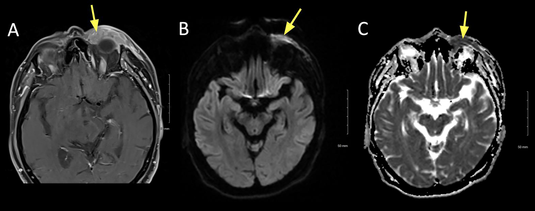
Figure 4:

A 79 year old female presented with slowly enlarging left upper and lower eyelid lesions over the past year. Diffusion weighted imaging (DWI) showed hyperintense signal, suggesting a hypercellular lesion. Biopsy confirmed mucosa-associated lymphoid tissue (MALT) lymphoma.
A. Axial T1+C FS at the superior lid level showing uniform contrast enhancement of the left upper lid (arrow).
B. Axial DWI showing hyperintense signal in the left upper lid.
C. ADC map of the lesion showing corresponding hypointense signal, signifying true restricted diffusion.
