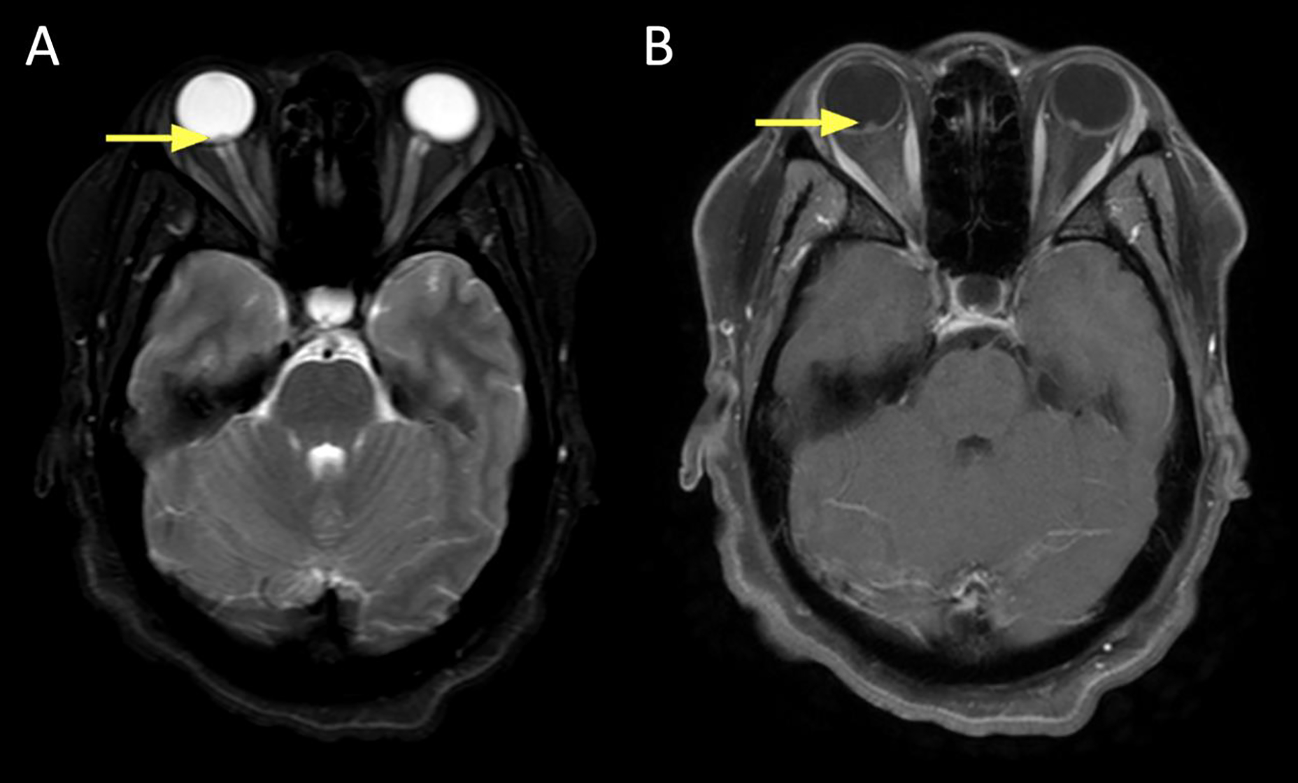
Figure 6:

A 41 year old male presented with left-sided ptosis, dysmotility, and orbital mass causing compressive optic neuropathy. Biopsy was consistent with granular cell tumor.
A. Axial T1+C FS at the mid/upper orbit reveals avid and uniform contrast enhancement of the lesion, which marginates the left medial rectus (yellow).
B. Axial DWI revealing intermediate signal in the lesion (arrow).
C. ADC map revealing slightly low signal in the lesion (arrow).
