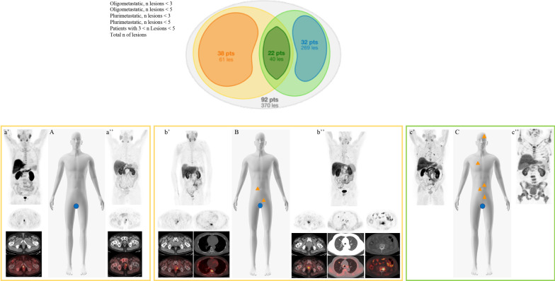
Fig. 1.
Schematic representation of the final classification in oligometastatic and plurimetastatic patients based on the number of lesions identified at [18F]FMCH (up to 3/5 and > 3/5, respectively) PET/CT findings at analysed population (upper panel). At lower panel clinical examples from the series: schematic representation (A–C) and [18F]FMCH PET/CT findings (a’–a’’, b’–b’’, c’–c’’, upper panel MIP images and lower panel top-down transaxial emission, CT and superimposed PET/CT at different levels) of oligorecurrent disease (A, a’, a’’), oligometastatic patients (B, b’, b’’) and multimetastatic patient (C, c’ and c’’)

