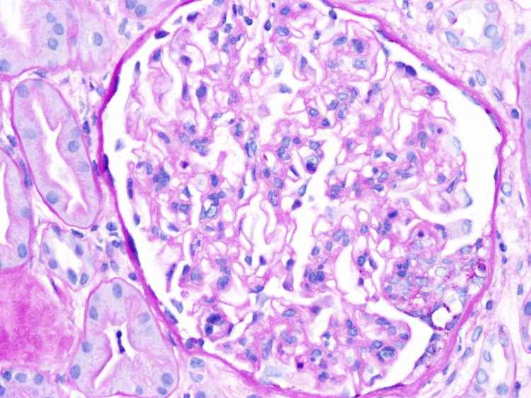
Figure 1. The light microscopy shows diffuse but variable glomerular mesangial matrix expansion along with diffuse moderate mesangial cell proliferation and focal endocapillary cell proliferation.
The tubules show a focal dilatation along with epithelial cell injury, necrosis, or flattening. The interstitium demonstrates moderate interstitial fibrosis with no significant interstitial inflammation.

