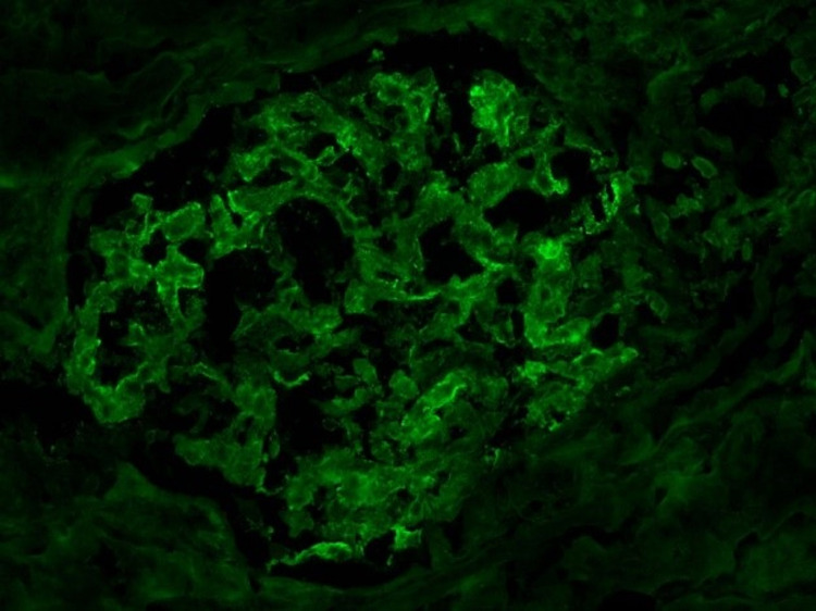
Figure 2. The immunofluorescence analysis demonstrates trace-to-mild diffuse granular glomerular mesangial IgA deposition along with trace IgM and C3 staining.
No linear C3 staining of the tubular membranes is apparent. The lambda and kappa light chain staining are heterogeneous (i.e., non-monoclonal) in the IgA staining pattern and involve tubular casts. No significant staining is apparent for IgG, C1q, or fibrinogen.

