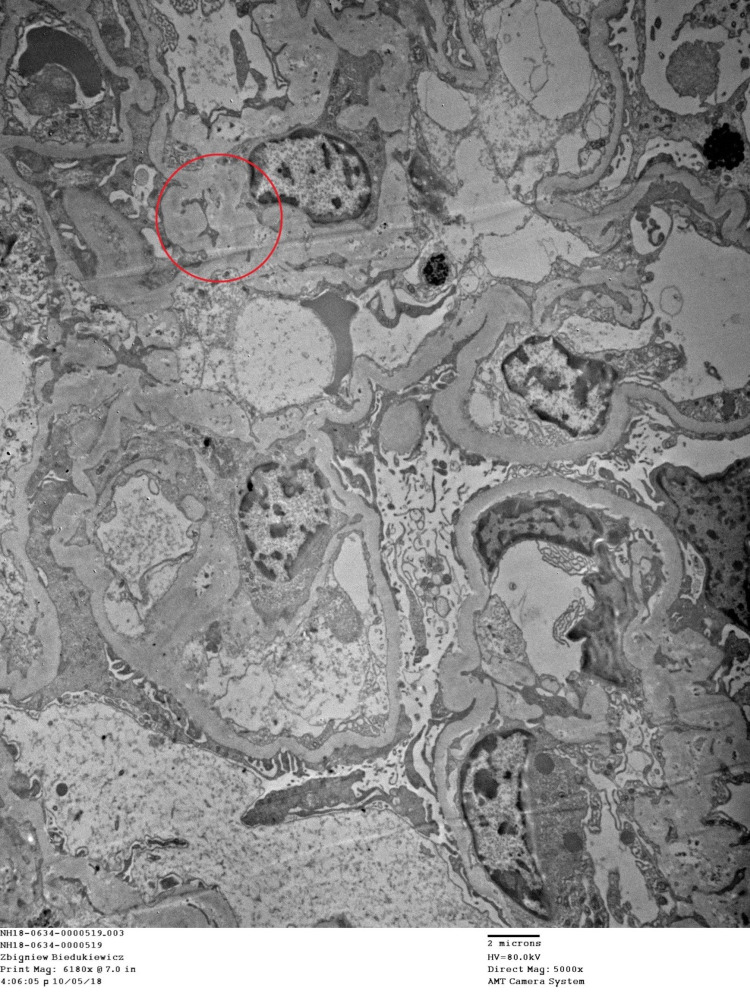
Figure 3. Electron microscopy demonstrates a viable glomerulus with variable mesangial matrix expansion and glomerular basement membrane thickening but no lamination or defects.
There are small mesangial deposits apparent. A fibro-cellular glomerular crescent is apparent. Glomerular foot process effacement is segmentally present. The tubules display epithelial cell changes consistent with acute tubular injury.

