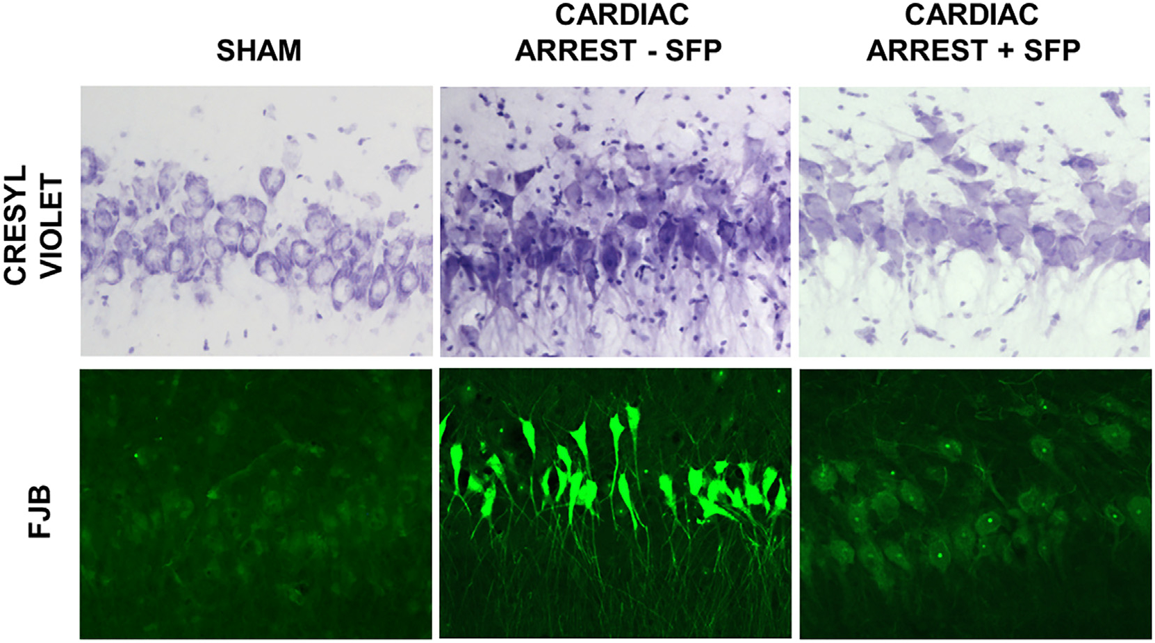
Fig. 2.

Hippocampal neuronal death at 24 hr after cardiac arrest and protection by sulforaphane. Canine brain sections were stained with cresyl violet for morphologic alterations and with Fluoro Jade B (FJB) for neuron-specific damage and death. Following Sham cardiac arrest, hippocampal CA1 neurons appeared healthy with homogeneous discrete nuclei and cell as shown with cresyl violet-stained sections. Following cardiac arrest and 24 hr resuscitation, most neurons displayed dark, pyknotic nuclei and were surrounded by cell debris. Sulforaphane administration reduced but did not eliminate neuronal abnormalities. Sections stained with Fluoro Jade B exhibited little staining in Shams and in dogs following cardiac arrest and treatment with sulforaphane. Fluoro Jade B was much greater in animals that underwent cardiac arrest with no sulforaphane treatment.
