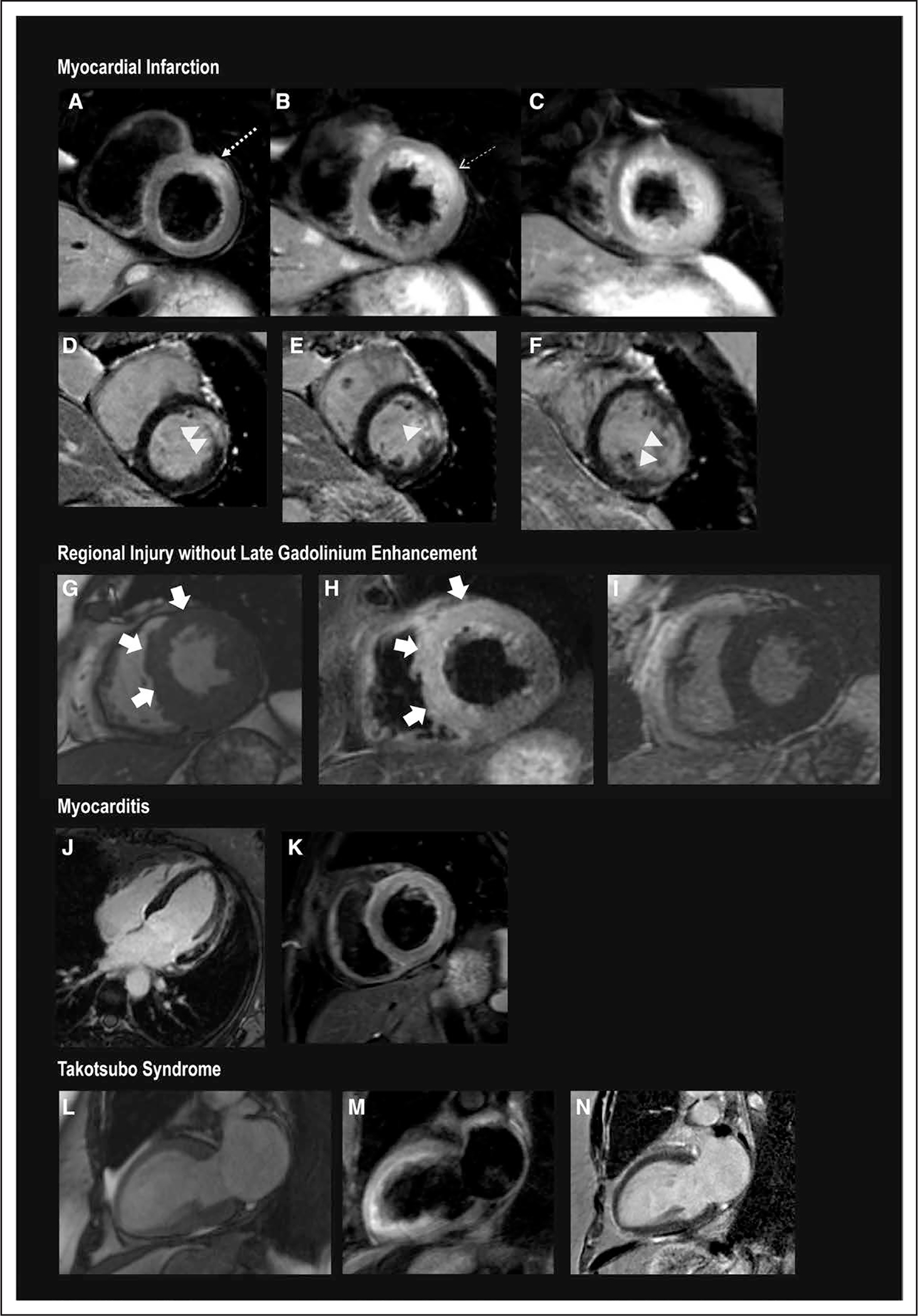
Figure 4. Representative CMR images in women with MINOCA.

A through F, Myocardial infarction. A through C, T2-weighted images. D through F, Late gadolinium enhanced images, arranged from basal (left) to apical (right). There is subendocardial-to-transmural late gadolinium enhancement of the basal anterior and anteroseptal, mid anterior, and the apical anterolateral wall (>). T2-weighted imaging demonstrates increased signal within and extending beyond the area of late gadolinium enhancement (—→), affecting the LAD territory, including all apical segments. G through I, Regional edema without late gadolinium enhancement. The anterior and anteroseptal walls were hypokinetic (C, systolic frame). This wall motion abnormality was matched by evidence of T2 signal enhancement in the matching regions (D). There was no evidence of infarction on late gadolinium-enhanced imaging (E). J and K, Myocarditis. Multiple foci of late gadolinium enhancement of the anterolateral wall (F), which is matched by T2 hyperintensity in the matching anterolateral wall and anterior wall (G). On cine imaging, there was diffuse hypokinesis of the left ventricle. L and M, takotsubo syndrome. H, Systolic frame demonstrates hypokinesis of the anterior wall, apex, and apical-to-mid inferior wall with preserved contraction at the base. I, T2-weighted imaging with hyperintensity in the anterior wall, apex, and apical-to-mid inferior wall. N, Absence of late gadolinium enhancement. CMR indicates cardiac magnetic resonance; LAD, left anterior descending; and MINOCA, myocardial infarction with nonobstructive coronary arteries (<50% stenosis in all major epicardial vessels).
