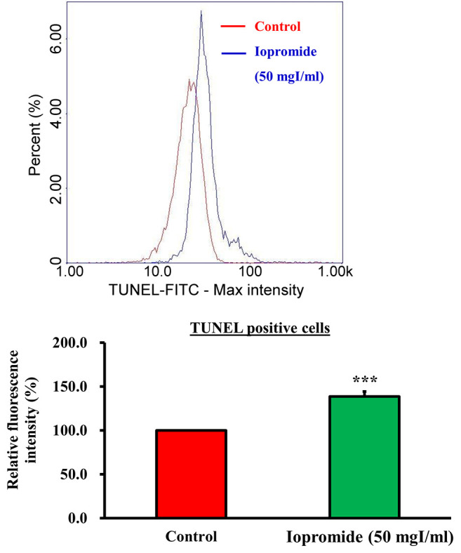
Figure 3. Iopromide induces DNA fragmentation and apoptosis. (A) HEK 293 cells were incubated with 50 mgI/ml iopromide for 48 h. DNA breaks were determined using the TUNEL assay. TUNEL-positive cells were analyzed using the NucleoCounter® NC-3000™ advanced image cytometer. (B) The relative fluorescence intensity was calculated using the ratio of the average mean of sample fluorescence intensity of iporomide-treated cells to that of untreated control cells. The relative fluorescence intensity is expressed as fold change. The results are expressed relative to those of the untreated control and values are presented as mean±SE (n=3) (***p<0.001).

