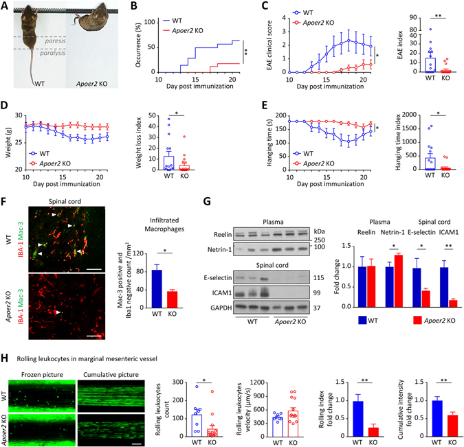
Figure 1. ApoER2 deletion protects mice from EAE by reducing leukocyte recruitment.
A, EAE was induced by MOG immunization in 8 weeks old ApoER2 WT (n=14) and KO (n=17) male littermates (the experiment was performed twice and the results were pooled). B, Disease incidence (clinical signs of paralysis) was monitored daily for 21 days. C-E, EAE severity was evaluated daily from day 10 using (C) an EAE clinical score (from 0=unaffected to 10=dead), (D) weight loss and (E) inverted hanging test (for a maximum time of 180 sec). C-E, EAE severity, weight loss and hanging time indices were calculated for each animal by integrating daily scores over the course of the experiment. F, In the spinal cord, the total number of infiltrating macrophages (Mac-3-positive, Iba-1-negative; indicated by the arrows) was evaluated by immunofluorescence (scale = 50 μm). G, 21 days after EAE induction, expression of Reelin and Netrin-1 in the plasma, as well as E-selectin and ICAM-1 in the spinal cord were measured by immunoblotting. H, 4-week-old ApoER2 WT (n=8) and KO (n=14) male and female littermates received Rhodamine 6G by retro-orbital injection to label circulating leukocytes. Next, intravital microscopy was performed to record numbers and speed of rolling leucocytes on marginal mesenteric vessels. 6 different vessels/mouse were recorded for 10 sec and analyzed; cumulative pictures represent individual images (100 frames/second) integrated over a 10 sec period and stacked; rolling index = leucocyte number / velocity. *p<0.05 and **p<0.01 (Mantel-Cox test, 2-way ANOVA or t-test / Mann Whitney).

