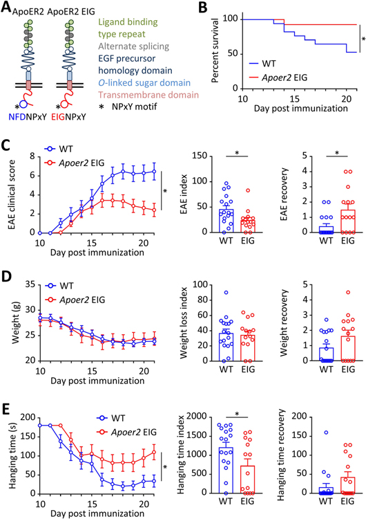
Figure 2. Impaired ApoER2 function in mice reduces EAE severity.
A, We used ApoER2-EIG mutant mice in which the ApoER2 NFDNPVY docking motif for Dab1/2 was rendered non-functional to disrupt the ApoER2-Dab interaction, thereby impairing ApoER2 signaling and function. This line was crossed to Cx3cr1-GFP mice expressing GFP in monocytes to follow the infiltration of this specific inflammatory cell population. B-G, EAE was induced by MOG immunization in 8 weeks old ApoER2 WT (n=17) and EIG (n=14) male littermates. B-E, EAE severity was scored daily starting at day 10 by recording (B) survival, (C) EAE clinical score (from 0=unaffected to 10=dead), (D) weight loss and (E) inverted grid hanging endurance (for a maximum time of 180 sec). C-E, EAE severity, weight loss and hanging time indices are calculated for each animal by integrating daily scores over the course of the experiment. *p<0.05 and **p<0.01 (Mantel-Cox test, 2-way ANOVA or t-test / Mann Whitney).

