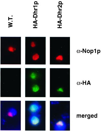
FIG. 3.
Dhr1p and Dhr2p localize to the nucleolus. Indirect immunofluorescence on cells expressing an HA epitope fusion of either Dhr1p or Dhr2p. The Dhr fusions were detected with a rat anti-HA (α-HA) monoclonal antibody, followed by FITC staining. For Nop1p detection, a mouse anti-Nop1p (α-Nop1p) monoclonal antibody was used in combination with Cy3 staining. Both HA fusions showed an intranuclear staining (green channel) which colocalized with Nop1p (red channel) and revealed a classical nucleolar crescent-like shape structure (yellow in merged images). DNA was stained with DAPI (blue in merged images). W.T., wild type.

