FIGURE 4.
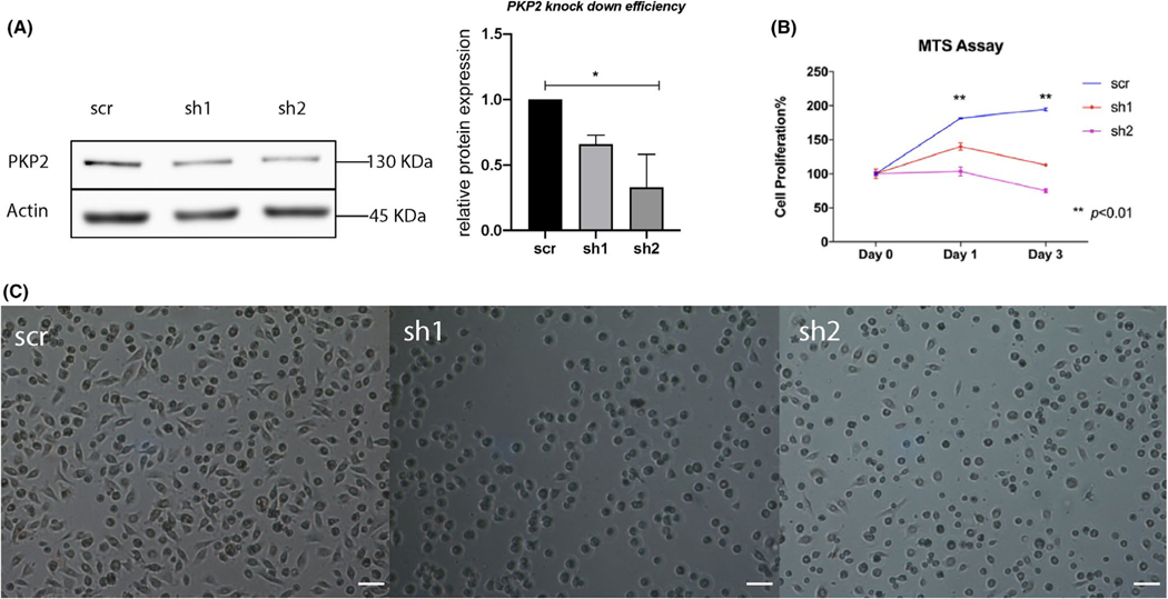
PKP2 deficiency impairs cell proliferation and spreading. (A) PKP2 was moderately knocked down by short-hairpin 1 (sh1) and short-hairpin 2 (sh2) lentivirus-shRNA constructs in HGEPs compared to scramble control (scr), as shown by western blotting. Densitometry analysis of PKP2 protein expression in sh1 and sh2 from two separate western blots (right panel) is to demonstrate the knockdown efficiency. (B) Epithelial cells with deficient PKP2 displayed significantly inhibited cell proliferations. ** indicates p < .01. Error bars represent standard deviations. 2 × 10⁴ puromycin-selected cells had been seeded on 96-well plates and waited for 48 h before we collected culture medium at day 0 (the first medium collection), 1, and 3 for MTS assay. (C) HGEPs with deficient PKP2 displayed impaired cell spreading 1 h after being seeded. Scale bar is 31 μm

