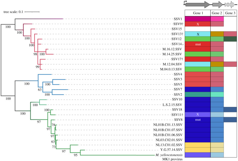
Figure 3.
Diversity of toxin genes found among representative SSVs. Maximum-likelihood core gene tree of a subset of SSVs representing the diversity of viruses that have been isolated and sequenced to date; branches with greater than 50% bootstrap support are labelled. Branch colour represents geographical location that viruses were isolated from: Japan (purple), Kamchatka, Russia (Uzon Geyser—dark red, Mutnovsky Volcano—light red), Iceland (blue) or United States (Lassen National Park—light green, Yellowstone National Park—dark green). SSVs labelled with † have been found to confer a toxic phenotype; that marked with ○ does not [17]. Putative toxin genes upstream of the VP4 tail fibre gene were clustered using CD-HIT based on 50% identity; clusters are represented by the different colours in the table to the right of the tree. Genes with an ‘X’ were tested in this work; those marked as ‘mut’ contain a mutation that would suggest the toxin gene is non-functional, such as a mutated start codon in Gene 1 of SSV14 or a frameshift in Gene 1 of SSV8.

