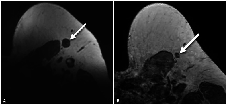
Fig. 2. A-47-year-old female with right breast cancer.
A, B. Axial T1-weighted MR image performed after recent COVID-19 vaccination shows enlarged left axillary node (arrow, A). The same node had a normal appearance prior to vaccine administration with visible fatty hilum on axial T1-weighted MR image obtained 54 days earlier (arrow, B).

