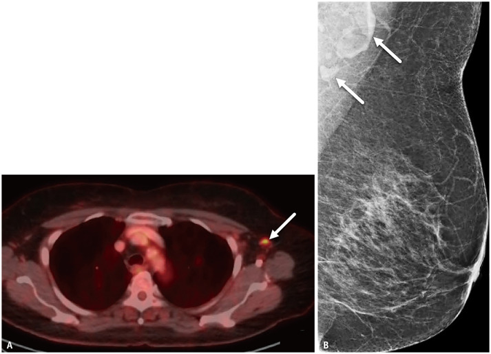
Fig. 6. A-76-year-old female with history of lymphoma.
A. Axial fused PET-CT image performed 15 days after 1st dose vaccine demonstrates normal sized left axillary node with increased fluorodeoxyglucose uptake (arrow). B. Left axillary nodes (arrows) appear normal in morphology and stable since prior mammography.

