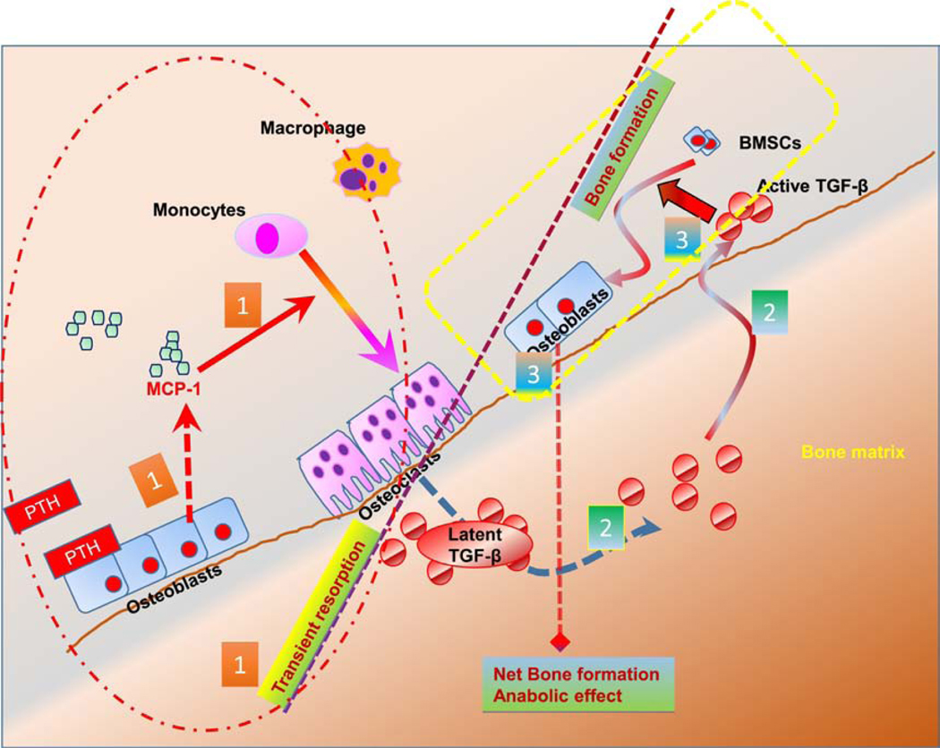
Figure 8: Schematic diagram of MCP-1 signaling and PTH mediated bone anabolic effects.
(1) Intermittent PTH rapidly increases MCP-1 expression in the osteoblast. Osteoblastic MCP-1 is necessary for recruitment of monocytes to macrophages and osteoclasts after PTH treatment and the latter’s transient enhanced breakdown of bone. (2) The bone matrix contains abundant latent TGF-β. Short-term osteoclastogenesis increases the release of active TGF-β from bone matrix. (3) Finally, the active TGF-β then acts on bone marrow stromal cells (BMSCs) and recruits them to osteoclastic resorption sites where they initiate new bone formation.

