
Extended Data Figure 7.
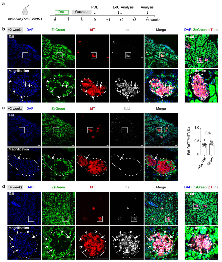
β cells are not generated from non-β cells after pancreatic ductal ligation.
(a) A schematic diagram illustrating experimental strategy of Dox induction, pancreatic ductal ligation (PDL) and analysis. (b) Immunostaining for tdT, ZsGreen and Ins on pancreatic tail (left) and head (right) sections collected after PDL 2 weeks. Arrows indicate tdT+Ins+ β cells. Arrowheads indicate zsGreen+Ins− non-β cells. (c) Immunostaining for tdT, ZsGreen and EdU collected after PDL. Arrowheads indicate EdU+tdT+ cells. EdU was injected 12 hours before sacrifice. Right panel shows quantification of the percentage of tdT+ cells with incorporated EdU. Numbers of investigated mice were as follows: PDL group, n = 5; Sham group, n = 5, Data are mean ± SEM, n.s., not significant (unpaired, two-sided, Student’s t-test). (d) Immunostaining for tdT, ZsGreen and Ins on pancreatic tail (left) and head (right) sections collected after PDL 4 weeks. Arrows indicate tdT+Ins+ β cells. Arrowheads indicate zsGreen+Ins− non-β cells. Scale bars, 100 μm. Each image is representative of 5 individual samples.