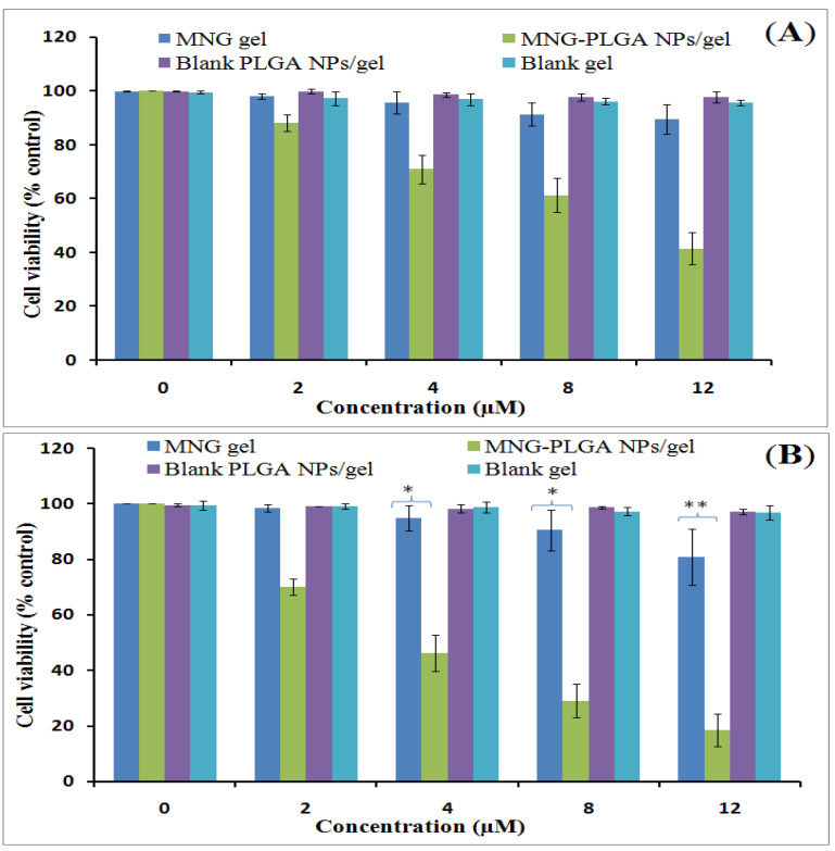
Figure 9.
Cell viabilitystudy of α-MNG gel, α-MNG-PLGA NPs gel, blank PLGA NPs gel and blank gel at concentrations, (0, 2, 4, 8, and 12 µM) after incubation times at 24 h (A); and after 48 h (B) in skin cancer cell line. Data indicated as mean ± SD (n = 3); (* p < 0.05), (** p < 0.01) compared with α-MNG gel.

