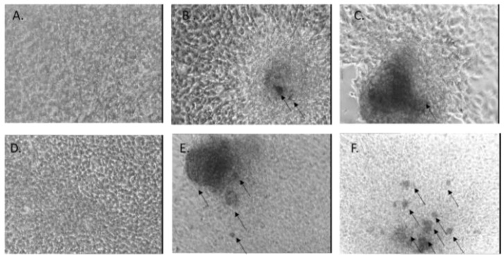
Figure 14.
Alizarin Red staining of MG-63 cells grown in normal culture conditions (control) for 7 days (A) or 14 days (D), or in the presence of alginate samples for 7 days (B,C) or 14 days (E,F). Images were obtained using an inverted phase-contrast microscope with a black-and-white camera attached. Dark areas correspond to Alizarin Red staining.

