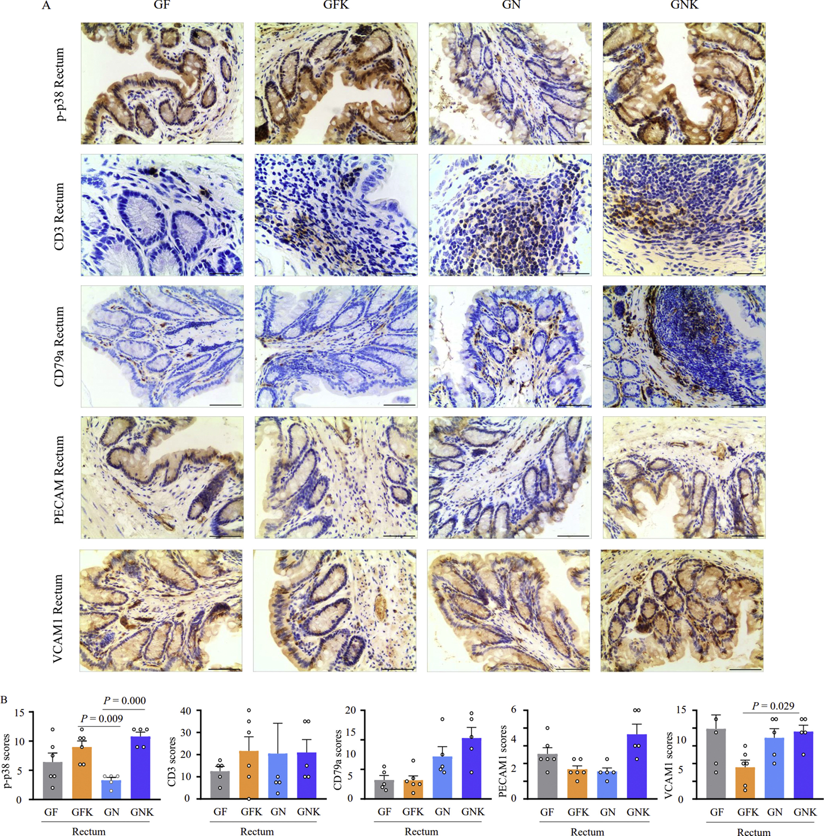
Fig. 5.

Immunohistochemical staining and inflammatory marker scoring of rectum tissue treated with KCO and KCO degrading bacteria. A: Representative images of rectal tissue samples stained for p-p38, CD3, CD79a, PECAM1, and VCAM1 inflammatory markers. Scale bar, 200 μm. B: Histopathology scores indicate inflammation severity in the rectum based on quantification of IHC staining results by ImageJ. Results are presented as mean ± SEM (n = 6). Tissue samples were collected from GF (germ free control group), GFK (5% KCO), GN (5 × 10⁸ CFU of B. xylanisolvens and E. coli), and GNK (5% KCO plus 5 × 10⁸ CFU of B. xylanisolvens and E. coli) treatment groups in germ-free Kunming mice. SEM, standard error of the mean.
