Figure 1.
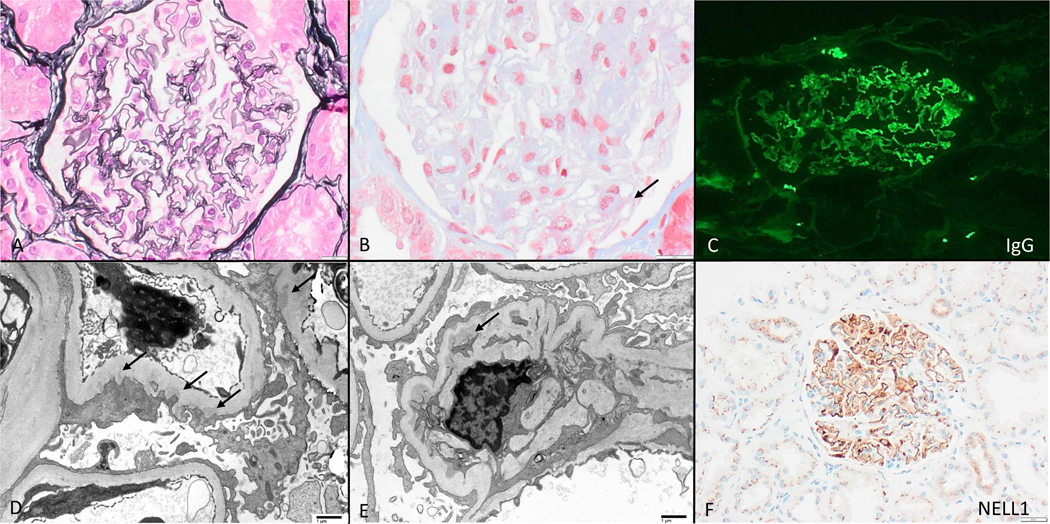
NELL1- associated membranous nephropathy in the setting of lipoic acid supplementation, with nearly-normal appearing glomeruli by light microscopy (A, Jones 400x) with subtle fuschinophilic subepithelial immune deposits (B, arrow, trichrome stain 600x). All cases had segmental capillary wall deposits of polyclonal IgG (C) which was often IgG1 dominant, and associated irregularly distributed subepithelial immune deposits by electron microscopy (D, arrows, direct magnification 2900x); mesangial deposits were present in 1 case (E, arrow, direct magnification 2900x). Glomerular deposits reacted with NELL 1 (F, 200x).

