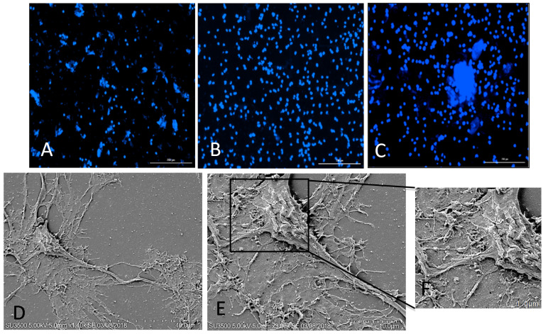
Figure 2.
Attachment of rNSC cells to the CNC–, PDL–, and CNC–Lys–coated surfaces and SEM images of differentiated rat NPCs on the CNC–Lys surface. The plated NSCs attached well to the (A) CNC, (B) PDL, and (C) CNC–Lys surfaces, Scale bar: 200 μm. Nuclei were counterstained with DAPI. (D) Fully differentiated neurons on the CNC–Lys surface. Scale bar: 40 μm. (E) Cell body of neurons with neurites, shown in more detail. Scale bar: 10 μm. (F) High magnification of the boxed region, showing details of the surface of the cell body and its interaction with the substratum. Scale bar: 1 μm.

