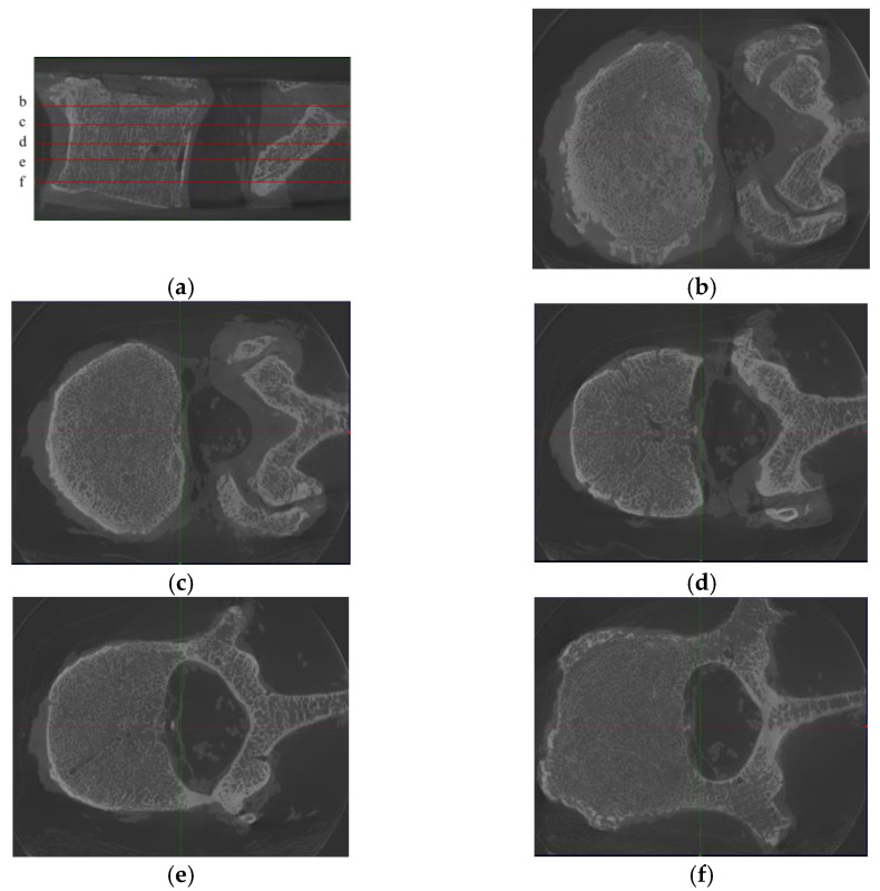
Figure 2.
Samples from one L2 vertebra, where (a) is the sagittal position image of the L2 vertebra, with 5 noted slices (named b–f), and (b)–(f) are the corresponding axis position images. We found that although the images were from the same vertebra, the differences between the images of different slices were substantially large. Moreover, since the technique used in this paper is an image-to-image technique, there is no longer a holistic concept of “vertebra” in the training process but only discrete images.

