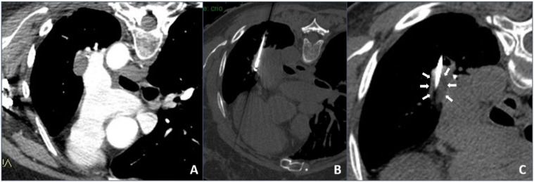
Figure 2.
Cryoablation procedure: (A) contrast-enhanced axial CT scan showing a left hilum neoplastic lesion adjacent to the left pulmonary artery; (B) intra-procedural unenhanced axial CT scan with maximum intensity projection (MIP) reconstruction showing the cryoablation needle inside the lesion; (C) intra-procedural unenhanced axial CT scan with MIP reconstruction showing direct visualization of the hypodense core necrotic volume.

