
Figure 1: Introduction to Clytia.
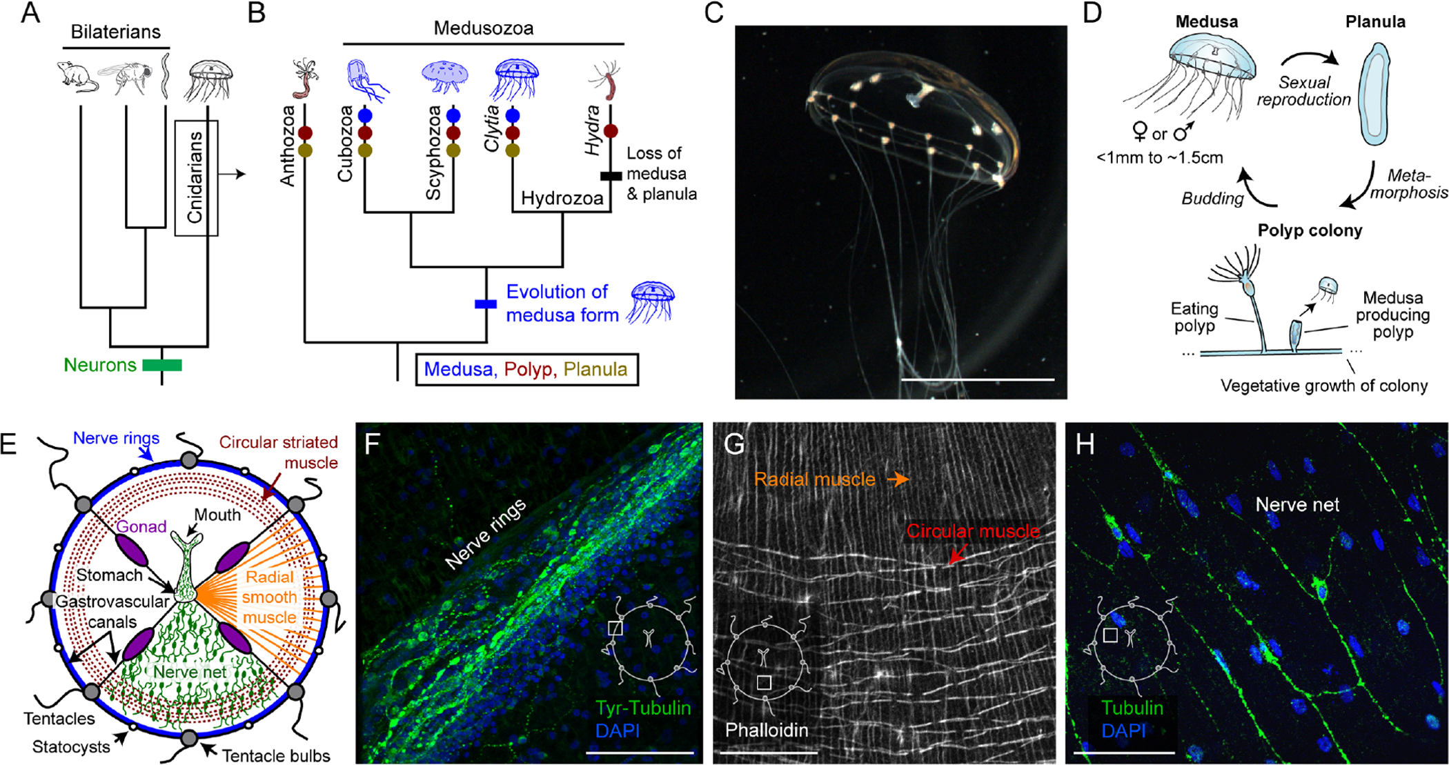
(A) Relationship between cnidarians, bilaterian lineages, and the appearance of neurons.
(B) Specific cnidarian lineages and transitions in body form. Text: taxonomic class; italics: genus.
(C) Clytia medusa. Scale: ~0.5cm.
(D) Clytia life cycle: medusa (jellyfish), planula (larva), and polyp stages.
(E) Medusa anatomy. Nerve net and radial muscle are shown in only one quadrant for clarity.
(F) Nerve rings. Green: anti-Tyrosinated Tubulin. Blue: DAPI. Scale: 50μm. Inset here and in G-H indicates anatomical location.
(G) Radial vs. circular muscle visualized with phalloidin. Scale: 50μm.
(H) Nerve net. Green: anti-aTubulin. Blue: DAPI. Scale: 50μm.