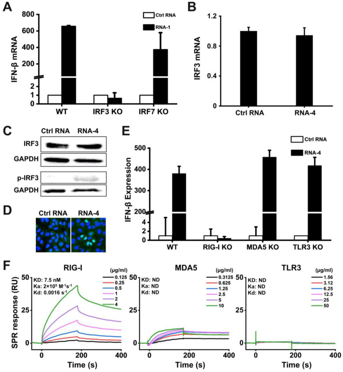
Figure 3. Immunostimulatory RNAs induce IFN-I production through RIG-I-IRF3 pathway.
(A) Wild-type (WT) HAP1 cells, IRF3 knockout HAP1 cells, or IRF7 knockout HAP1 cells were transfected with RNA-1 or scrambled RNA control for 48 h, and IFN-β mRNA levels were quantified by qPCR. Data are shown as fold change relative to the scrambled RNA control (N = 3). Note that IRF3 knockdown completely abolished the IFN-β response. (B) IRF3 mRNA levels measured in A549 cells transfected with immunostimulatory RNA-4 or a scrambled RNA control, as determined by qPCR and 48 h post-transfection (data are shown as fold change relative to the control RNA; N = 3). (C) Total IRF3 protein and phosphorylated IRF3 detected in A549 cells transfected with RNA-4 or scrambled RNA control at 48 h post transfection as detected by Western blot analysis (GAPDH was used as a loading control). (D) Immunofluorescence micrographs showing the distribution of phosphorylated IRF3 in A549 cells transfected with RNA-4 or scrambled RNA control at 48 h post transfection (Green, phosphorylated IRF3; blue, DAPI-stained nuclei; arrowheads, nuclei expressing phosphorylated IRF3). (E) Wild-type (WT) A549-Dual cells, RIG-I knockout A549-Dual cells, MDA5 knockout A549-Dual cells, or TLR3 knockout A549 cells were transfected with immunostimulatory RNA-4 or a scrambles RNA control and 48 h later, IFN-β expression levels were quantified using the Quanti-Luc assay or qPCR (data are shown as fold change relative to the scrambled RNA control; N = 6). Note that RIG-I knockout abolished the ability of the immunostimulatory RNAs to induce IFN-β. (F) SPR characterization of the binding affinity between cellular RNA sensors (RIG-I, MDA5, and TLR3) and RNA-1, which were immobilized on a streptavidin (SA) sensor chip. Equilibrium dissociation constant (KD), association rate constant (Ka), and dissociation rate constant (Kd) are labeled on the graphs.

