FIG. 1.
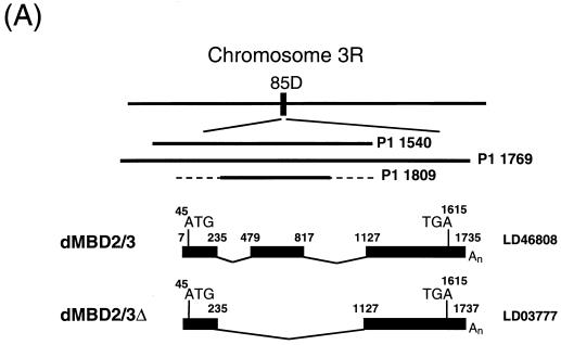
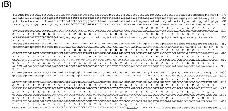
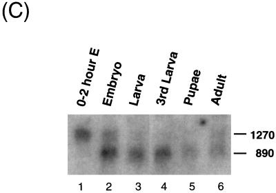
Structure and expression of the Drosophila dMBD2/3 gene. (A) Genomic and cDNA organization of dMBD2/3. The chromosomal location (85D) of the gene and the three P1 clones spanning the region are indicated. Shown below the P1 clones are the cDNAs for dMBD2/3 and dMBD2/3Δ, which were derived from the cDNA clones LD46808 and LD03777, respectively. The base numbering of the two cDNAs corresponds to that of the genomic sequence of dMBD2/3 shown in panel B. (B) Genomic sequence of dMBD2/3. The sequence of the dMBD2/3 gene was deduced from the three P1 clones. The putative site of transcriptional initiation, as inferred from the sequences of the two cDNA clones, is designated +1. The two introns and the regions upstream and downstream of the gene are shown with lowercase letters. The corresponding amino acids are also shown, with those homologous to the conserved amino acids among the vertebrate MBDs indicated by bold letters. The putative TATA box in the upstream promoter region and the poly(A) signal, AATAAA, are underlined. (C) Expression of dMBD2/3 and dMBD2/3Δ mRNA. Drosophila poly(A) RNAs isolated from different stages of development were analyzed by Northern blot hybridization with the cDNA clone LD03777 as the probe. The two transcripts (1,270 and 890 nt long) are indicated.


