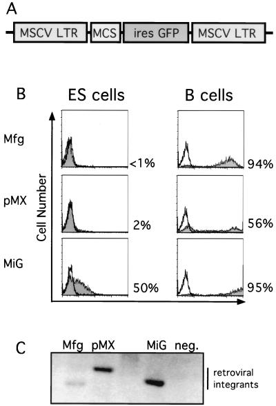
FIG. 1.
Efficient retroviral infection of ES cells. (A) Schematic diagram of MiG vector containing the MSCV LTR followed by a multiple cloning site (MCS) and an IRES-GFP cassette. (B) MSCV-based (MiG) but not Moloney virus-based (Mfg and pMX) retroviruses express in ES cells. B cells or ES cells were infected by the indicated retroviruses and assayed by flow cytometry 2 days postinfection. Uninfected cells (unshaded) and infected cells (shaded area) were electronically gated for live cells and subsequently analyzed for GFP fluorescence and for cell number. Percentages of GFP-positive cells are indicated. (C) Comparable levels of integration of different retroviruses into ES cells, determined by Southern blot analysis of genomic DNA purified from infected and uninfected ES cells 2 days postinfection, digested with KpnI, a restriction site present within the LTRs, and probed with the GFP coding sequence.

