Figure 3.

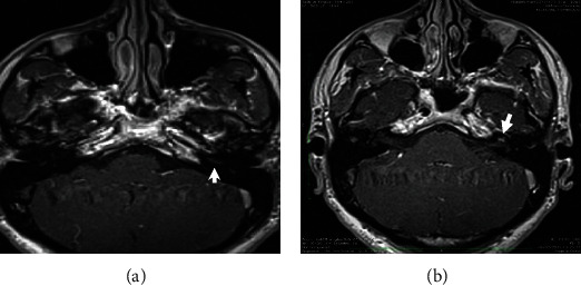
Magnetic resonance imaging of the temporal bone: axial postcontrast T1-weighted slices showing no enhancement after gadolinium injection in the basal (a) and the second (b) turns of the left cochlea.

Magnetic resonance imaging of the temporal bone: axial postcontrast T1-weighted slices showing no enhancement after gadolinium injection in the basal (a) and the second (b) turns of the left cochlea.