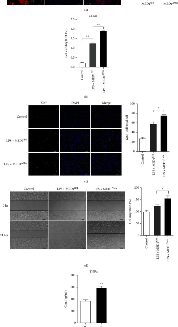
Figure 5.

MED1 deficiency in macrophages induces more migration and proliferation of vascular smooth muscle cells. (a) Representative immunofluorescence staining and quantification of α-smooth muscle actin (SMA) in injured arteries (n = 4–5). (b–e) After 24 h of LPS treatment, the culture medium of macrophages was collected and centrifuged to remove suspended cells for subsequent experiments. (b) The proliferation of smooth muscle cells was measured by CCK8 assay after adding DMEM high glucose medium (control), MED1 knockout macrophage medium (LPS+MED1ΔMac), or MED1fl/fl macrophage medium (LPS+MED1fl/fl) for 24 h. (c) Representative immunofluorescence staining and quantification of Ki67 in smooth muscle cells after adding DMEM high-glucose medium (control), MED1 knockout macrophage medium (LPS+MED1ΔMac), or MED1fl/fl macrophage medium (LPS+MED1fl/fl) for 24 h. (d) Migration of smooth muscle cells was determined by wound healing after adding DMEM high glucose medium (control), MED1 knockout macrophage medium (LPS+MED1ΔMac), or MED1fl/fl macrophage medium (LPS+MED1fl/fl) for 24 h. (e) ELISA analysis of TNFα levels in culture medium of LPS-treated MED1fl/fl macrophage (LPS+MED1fl/fl) or MED1ΔMac macrophage (LPS+MED1ΔMac) (n = 4). The data are expressed as the mean ± SEM. ∗p < 0.5; ∗∗p < 0.01.
