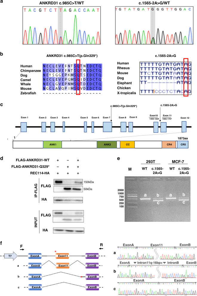
Fig. 2. ANKRD31 p.Gln329* impaired ANKRD31–REC114 interaction and c.1565-2A>G affected RNA splicing.
(a) Chromatograms of the two heterozygous variants in ANKRD31. (b) The variants were highly conserved among species. (c) ANKRD31 c.985C>T (p.Gln329*) localized in exon 7, before conserved region 5 (CR5), which was responsible for interaction with REC114. Splice site variant c.1565-2A>G localized at the donor splice site of intron 10. (d) Coimmunoprecipitation (Co-IP) analysis showed ANKRD31 p.Gln329* generated a truncated protein and impaired ANKRD31–REC114 interaction. (e) After 48 hours of transfection in two human cell lines (HEK293T and MCF-7 cell), agarose gel electrophoresis showed two belts of c.1565-2A>G transcripts in contrast with wild type. (f) Sequence analysis demonstrated that c.1565-2A>G variant caused two transcripts: belt b lacked exon 11 while contained 16 bp of intro11 and intron B; belt c skipped exon 11.

